Basic Ultrasound for Paediatrics
This Paediatric Ultrasound course is intended to give a strong foundation in using Point-of-Care Ultrasound (PoCUS) equipment, obtaining useful views and understanding the applications of PoCUS in the care of children in acute and emergency settings.
Next Date: 26th September 2024
Fee: £370 inc. VAT
Venue: Terracotta Court (ground floor), 167 Tower Bridge Road, London, SE1 3LN
19 years of experience in teaching RCEM courses
17 000 clinicians have taken our courses
4.9 out of 5 is our average Google review score
About Our Basic Paediatric Ultrasound Course
What Does the Course Cover?
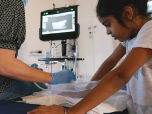
This course covers ultrasound equipment, probes and the most important controls (probe selection, presets, depth, gain, freeze, linear measurement, colour-flow Doppler and M-mode).
The topics covered include vascular access, lung ultrasound, basic cardiac ultrasound (ECHO), abdominal ultrasound including liver, kidney and bladder localisation and the identification of free fluid, foreign body localisation and nerve identification for local anaesthetic infiltration.
What Skills Will You Develop?
You will learn how to rapidly familiarise yourself with an ultrasound machine making the appropriate probe selection and instrument adjustments in order to obtain useful images.
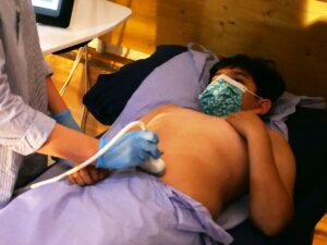
You will practice obtaining PoCUS images of the lungs, heart, abdominal organs, veins, nerves and foreign bodies.
By the end of this course, you will be able to handle a range of different ultrasound machines, including hand-held devices. You will understand the basis for different probe selection, how to adjust depth and gain and to use freeze, colour-flow Doppler and M-mode. You will understand how to optimise an ultrasound image. You will learn how to use ultrasound to guide a needle or cannula.
How Is the Course Taught?
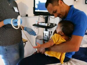
On the course, short lectures will be delivered covering the applications of PoCUS in the care of children in acute and emergency settings. The lectures will alternate with focused practical scanning sessions.
You will work with a scannable paediatric manikin, simulating a two-year-old, which has several pathologies including free fluid in the chest and abdomen, appendicitis, intussusception, and others. You will also practice scanning on healthy volunteers and simulators able to reproduce a wide range of lung, heart and abdominal pathologies.

